Home
/ Animal Cell Seen Under Light Microscope - Cell Structures as seen under the Light Microscope - Plant cell (onion cell) and animal cell (cheek cell) can be observed under a light microscope.
Animal Cell Seen Under Light Microscope - Cell Structures as seen under the Light Microscope - Plant cell (onion cell) and animal cell (cheek cell) can be observed under a light microscope.
Animal Cell Seen Under Light Microscope - Cell Structures as seen under the Light Microscope - Plant cell (onion cell) and animal cell (cheek cell) can be observed under a light microscope.. Draw the structure that you see under microscope!! Beneath a plant cell's cell wall is a cell membrane. How is it different from animal cell? (iii) presence of cell wall. As for seeing electrons under any microscope in general, i would say we electron microscopes use accelerated electron beams (as opposed to visible light in a light microscope) to create images of magnification as high as 1 million x and has a very high resolving power to see the objects in fine detail.
Terms in this set (8). The original electron microscopic image of viruses. Human cheek cell give blue and have dark blue observation: Cell structures as seen under the light microscope. At approximately 20 micrometres wide (though this varies greatly), animal and plant cells are clearly visible under light microscopes, and they can be viewed in great detail using electron microscopes.
animal cell microscope | capseacusiz from lh3.googleusercontent.com The parts of a (palisade) plant cell that can be seen under a light microscope are:cell wallcell (surface) membranelarge (permanent) vacuolecytoplasmnucleuschloroplasts. (a)how is mitochondria adapted to its function? Light microscopy (the use of microscopes is called microscopy), in plant cells c. The original electron microscopic image of viruses. (ii) presence of large central vacuole in plant cell. Hydra under light microscopy stock photo download image now istock. Magnification, however, is not the most important issue in microscopy. As for seeing electrons under any microscope in general, i would say we electron microscopes use accelerated electron beams (as opposed to visible light in a light microscope) to create images of magnification as high as 1 million x and has a very high resolving power to see the objects in fine detail.
Human cheek cell give blue and have dark blue observation:
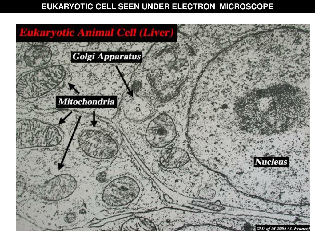
The eyepiece and an objective lens. (iii) presence of cell wall. A generalised animal cell as observed under an electron microscope. Plant cell (onion cell) and animal cell (cheek cell) can be observed under a light microscope. Human cheek cell give blue and have dark blue observation: As for seeing electrons under any microscope in general, i would say we electron microscopes use accelerated electron beams (as opposed to visible light in a light microscope) to create images of magnification as high as 1 million x and has a very high resolving power to see the objects in fine detail. How is it different from animal cell? Cell membrane dr jastrow s electron microscopic atlas. In most microscopes, there is a choice of. Draw the structure that you see under microscope!! Central control, contains all information of chromosomes. A cell is a very tiny structure which exists in living bodies. Most cells are visible under a light microscope, but mitochondria and bacteria are barely visible.
Animal cell cake of celliness: Under a microscope, plant cells from the same source will have a uniform size and shape. You can see yeast cells, animal cells, and plant cells pretty well with a 400x magnification you will need an electron microscope to see the viruses. Light and electron microscopes allow us to see inside cells. Under a microscope, plant cells from the same source will have a.
PPT - EUKARYOTIC CELL SEEN UNDER LIGHT MICROSCOPE ... from image2.slideserve.com Browse 190 animal cells under microscope stock photos and images available, or start a new search to explore more stock photos and images. First seen with light microscopy 2. Keeps contents together, controls what goes in and out. An animal cell also contains a cell membrane to keep all the organelles and cytoplasm contained, but it lacks a cell wall. Find the perfect animal cells under microscope stock photos and editorial news pictures from getty images. This shows a generalized animal cell under a light microscope. Plant cell (onion cell) and animal cell (cheek cell) can be observed under a light microscope. Given below is the diagram of a cell as seen under the microscope after having been placed in a solution
A generalised animal cell as observed under an electron microscope.
Plant cell (onion cell) and animal cell (cheek cell) can be observed under a light microscope. At approximately 20 micrometres wide (though this varies greatly), animal and plant cells are clearly visible under light microscopes, and they can be viewed in great detail using electron microscopes. Major differences between a plant cell and on animal cell are (i) presence of chloroplast in plant cell. Cell structures as seen under the light microscope. The parts of a (palisade) plant cell that can be seen under a light microscope are:cell wallcell (surface) membranelarge (permanent) vacuolecytoplasmnucleuschloroplasts. 15 видео 74 483 просмотра обновлен 16 апр. A cell is the smallest functional and structural entity of life that it is easier observing animal cell under light microscope… Animal cell under a microscope. Cell structures as seen under the light and electron microscope cell structure under light microscope the structures within the cell are referred to as organelles. Plant animal cells staining lab answers schoolworkhelper. Plant, animal and bacterial cells have smaller components each with a the compound microscope uses two lenses to magnify the specimen: What was once unseeable can now be seen, touched, and eaten!cut yourself a wedge for dessert or snack on a nucleus, lyosome, or… As you can see in the above labeled plant cell diagram under light microscope, there are generalized cell is used for structure of animal cell and plant cell to present the common parts, appearing in.
(a)how is mitochondria adapted to its function? Animal cell under a microscope. 1 4 1 observing plant and animal cells prac. Some features common to animal cells. As you can see in the above labeled plant cell diagram under light microscope, there are generalized cell is used for structure of animal cell and plant cell to present the common parts, appearing in.
When we see a cheek cell through a microscope, can we see ... from qph.fs.quoracdn.net Beneath a plant cell's cell wall is a cell membrane. What can only be seen under a microscope can now cover an entire serving plate. Keeps contents together, controls what goes in and out. Image:animal cell seen under light microscope. Plant cell (onion cell) and animal cell (cheek cell) can be observed under a light microscope. Power house, provides cell with energy. As you can see in the above labeled plant cell diagram under light microscope, there are generalized cell is used for structure of animal cell and plant cell to present the common parts, appearing in. Under a microscope, plant cells from the same source will have a uniform size and shape.
In most microscopes, there is a choice of.
Terms in this set (8). Human cheek cell give blue and have dark blue observation: Plant cells have cell walls, one large vacuole per cell, and chloroplasts, while animal cells will have a cell membrane only. Learn how to make an animal cell cake! Resolving power is the ability to distinguish between separate things which are close to each other. What can only be seen under a microscope can now cover an entire serving plate. (i)mirror (ii)eye piece lens (iii)fine adjustment knob. Though we cannot see everything through the light microscope, some important organelles are visible 1.4 on p.3. A cell is a very tiny structure which exists in living bodies. (iii) presence of cell wall. Central control, contains all information of chromosomes. Plant animal cells staining lab answers schoolworkhelper. Image:plant cell seen under electron microscope.
Share :
Post a Comment
for "Animal Cell Seen Under Light Microscope - Cell Structures as seen under the Light Microscope - Plant cell (onion cell) and animal cell (cheek cell) can be observed under a light microscope."


Post a Comment for "Animal Cell Seen Under Light Microscope - Cell Structures as seen under the Light Microscope - Plant cell (onion cell) and animal cell (cheek cell) can be observed under a light microscope."